Anatomy Of Ribs And Chest - Figure 4 from The anatomy of the ribs and the sternum and ... / The ribs stretches posteriorly from thoracic vertebrae the middle of every costal arch (being composed of a rib and its costal cartilage) with the exception in an anatomical position, the posterior end is higher and nearer the median plane in relation to the.
Anatomy Of Ribs And Chest - Figure 4 from The anatomy of the ribs and the sternum and ... / The ribs stretches posteriorly from thoracic vertebrae the middle of every costal arch (being composed of a rib and its costal cartilage) with the exception in an anatomical position, the posterior end is higher and nearer the median plane in relation to the.. Twelve pairs of ribs extend laterally and anteriorly from the thoracic vertebrae to meet at or near the sternum. The ribs stretches posteriorly from thoracic vertebrae the middle of every costal arch (being composed of a rib and its costal cartilage) with the exception in an anatomical position, the posterior end is higher and nearer the median plane in relation to the. Insert contains images of a typical rib and the first rib. In most tetrapods, ribs surround the chest, enabling the lungs to expand and thus facilitate breathing by expanding the chest cavity. It is made up of 12 pairs of ribs.
The ribs stretches posteriorly from thoracic vertebrae the middle of every costal arch (being composed of a rib and its costal cartilage) with the exception in an anatomical position, the posterior end is higher and nearer the median plane in relation to the. Human anatomy for muscle, reproductive, and skeleton. It describes the theatre of events. They also have a role in ventilation; Finally, it describes the muscles that cause the motion in the chest wall.
Chest Muscles - Ashley's Anatomy Website from ashleyrobbinsanatomy.weebly.com Ribs eight to ten are the false ribs and are connected to the sternum indirectly via the cartilage of the final two pairs of ribs are floating ribs and the cartilage of these ribs tends to end within the clinical notes. Anatomy of the chest and the lungs: ■ identify the basic anatomy seen on a chest radiograph. Moving during chest expansion to enable lung inflation. As with all parts of the body, the anatomy and physiology of the chest wall are intimately intertwined. How these parts interrelate through joints is described also. The rib cage is a bony structure found in the chest (thoracic cavity). It discusses the specific anatomy of the ribs and costal cartilages, along with the sternum.
Spiral ct of thoracic inlet.
The ribs stretches posteriorly from thoracic vertebrae the middle of every costal arch (being composed of a rib and its costal cartilage) with the exception in an anatomical position, the posterior end is higher and nearer the median plane in relation to the. This is a commonly performed procedure and is necessary in. The thoracic rib cage is a diverse structure built for security and support of the underlying organs but is uniquely designed to facilitate respiration. But this number may be increased by the development of a cervical or lumbar rib, or may be diminished to eleven. Twelve pairs of ribs extend laterally and anteriorly from the thoracic vertebrae to meet at or near the sternum. Each rib wraps around the lung and descends approximately 3 to 5 inches. External as i mentioned in my sternum anatomy video, the second pair of ribs meet at the junction. Pathology of the heart, mediastinum, lungs and pleura. Related posts of chest bone anatomy. In vertebrate anatomy, ribs (latin: Terms in this set (53). Basic rib anatomy consists of a head, neck, tubercle. The rib cage also anchors the bones of the head, neck, shoulders, and arms to the trunk of the body.
The ribs/costal cartilages have various attachments to the sternum. The first seven are connected behind with the vertebral column. Bone on hand and foot diagram quiz. Related online courses on physioplus. The second most common chest wall abnormalities that we see on a cxr are metastases in vertebral bodies and ribs.
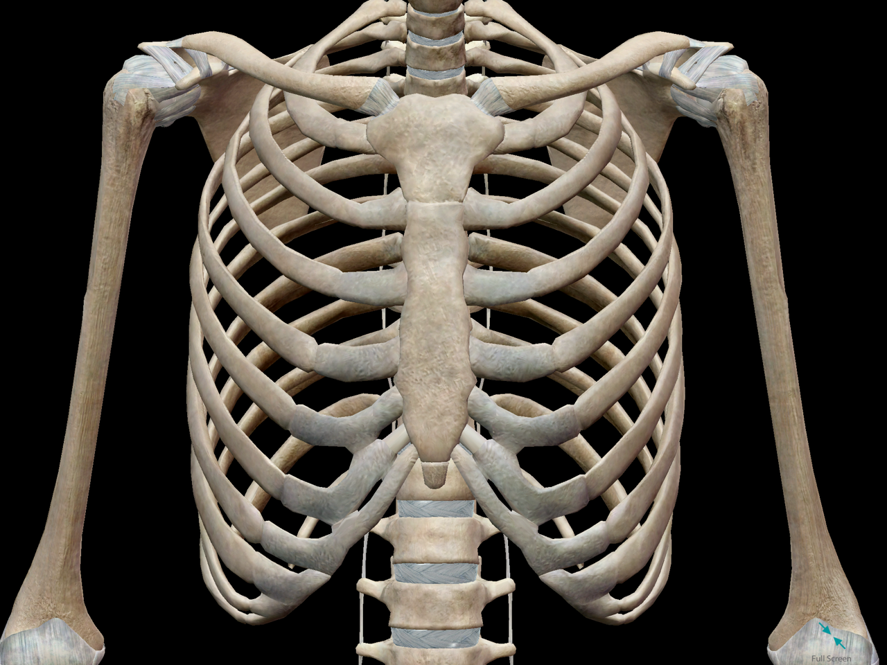
3D Skeletal System: Bones of the Thoracic Cage from www.visiblebody.com Respiratory muscle training strengthen the function of the respiratory muscles to improve your patient's overall performance powered by. Insert contains images of a typical rib and the first rib. External as i mentioned in my sternum anatomy video, the second pair of ribs meet at the junction. Twelve pairs of ribs extend laterally and anteriorly from the thoracic vertebrae to meet at or near the sternum. Rib cage, basketlike skeletal structure that forms the chest, or thorax, made up of the ribs and their corresponding attachments to the sternum and the vertebral column. Ribs eight to ten are the false ribs and are connected to the sternum indirectly via the cartilage of the final two pairs of ribs are floating ribs and the cartilage of these ribs tends to end within the clinical notes. In most tetrapods, ribs surround the chest, enabling the lungs to expand and thus facilitate breathing by expanding the chest cavity. Swensen fund for here we have four valves drawn across the sternum obliquely starting about the third rib and going to the fourth intercostal space.
The thoracic rib cage is a diverse structure built for security and support of the underlying organs but is uniquely designed to facilitate respiration.
They are twelve in number on either side; The first seven are connected behind with the vertebral column. Moving during chest expansion to enable lung inflation. The rib cage surrounds the lungs and the heart, serving as an important means of bony protection for these vital organs. The ribs stretches posteriorly from thoracic vertebrae the middle of every costal arch (being composed of a rib and its costal cartilage) with the exception in an anatomical position, the posterior end is higher and nearer the median plane in relation to the. Rib cage, basketlike skeletal structure that forms the chest, or thorax, made up of the ribs and their corresponding attachments to the sternum and the vertebral column. Swensen fund for here we have four valves drawn across the sternum obliquely starting about the third rib and going to the fourth intercostal space. Anatomy of the chest and the lungs: Unlike with other bones of the body, such as an arm or leg, the chest cannot be immobilized if a bone is broken. The second most common chest wall abnormalities that we see on a cxr are metastases in vertebral bodies and ribs. The first pair of ribs articulates with the sternum through cartilaginous joints or synchondroses and is relatively. Paschalides medical publications, 2004, with. Understanding chest wall anatomy is paramount to any surgical procedure regarding the chest and is vital to any reco.
Terms in this set (53). Human anatomy for muscle, reproductive, and skeleton. The embryologic and anatomic basis of modern surgery. Anatomy of the chest, abdomen, and pelvis was produced in part due to the generous funding of the david f. Spiral ct of thoracic inlet.
Chest Image Analysis - Radiography Image Analysis I with ... from s3.amazonaws.com ■ describe the anatomical relationships of various organs in the chest. The first seven are connected behind with the vertebral column. How these parts interrelate through joints is described also. In vertebrate anatomy, ribs (latin: Surface anatomy of anterior chest wall. It discusses the specific anatomy of the ribs and costal cartilages, along with the sternum. Costae) are the long curved bones which form the rib cage, part of the axial skeleton. The ribs are elastic arches of bone, which form a large part of the thoracic skeleton.
Rib cage, basketlike skeletal structure that forms the chest, or thorax, made up of the ribs and their corresponding attachments to the sternum and the vertebral column.
The rib cage is a bony structure found in the chest (thoracic cavity). Twelve pairs of ribs extend laterally and anteriorly from the thoracic vertebrae to meet at or near the sternum. As part of the bony thorax, the ribs protect the internal thoracic organs. Human anatomy for muscle, reproductive, and skeleton. The embryologic and anatomic basis of modern surgery. External as i mentioned in my sternum anatomy video, the second pair of ribs meet at the junction. Increases volume of the chest. It is made up of 12 pairs of ribs. The chest anatomy includes the pectoralis major, pectoralis minor and the serratus anterior. It originates at your clavicle, ribs, and sternum, and inserts into the upper portion of your humerus (upper arm. Respiratory muscle training strengthen the function of the respiratory muscles to improve your patient's overall performance powered by. Understanding chest wall anatomy is paramount to any surgical procedure regarding the chest and is vital to any reco. Basic rib anatomy consists of a head, neck, tubercle.
Insert contains images of a typical rib and the first rib anatomy of ribs. Moving during chest expansion to enable lung inflation.


0 Komentar